Pump-Priming Award: Interim Progress Report
1. Summary
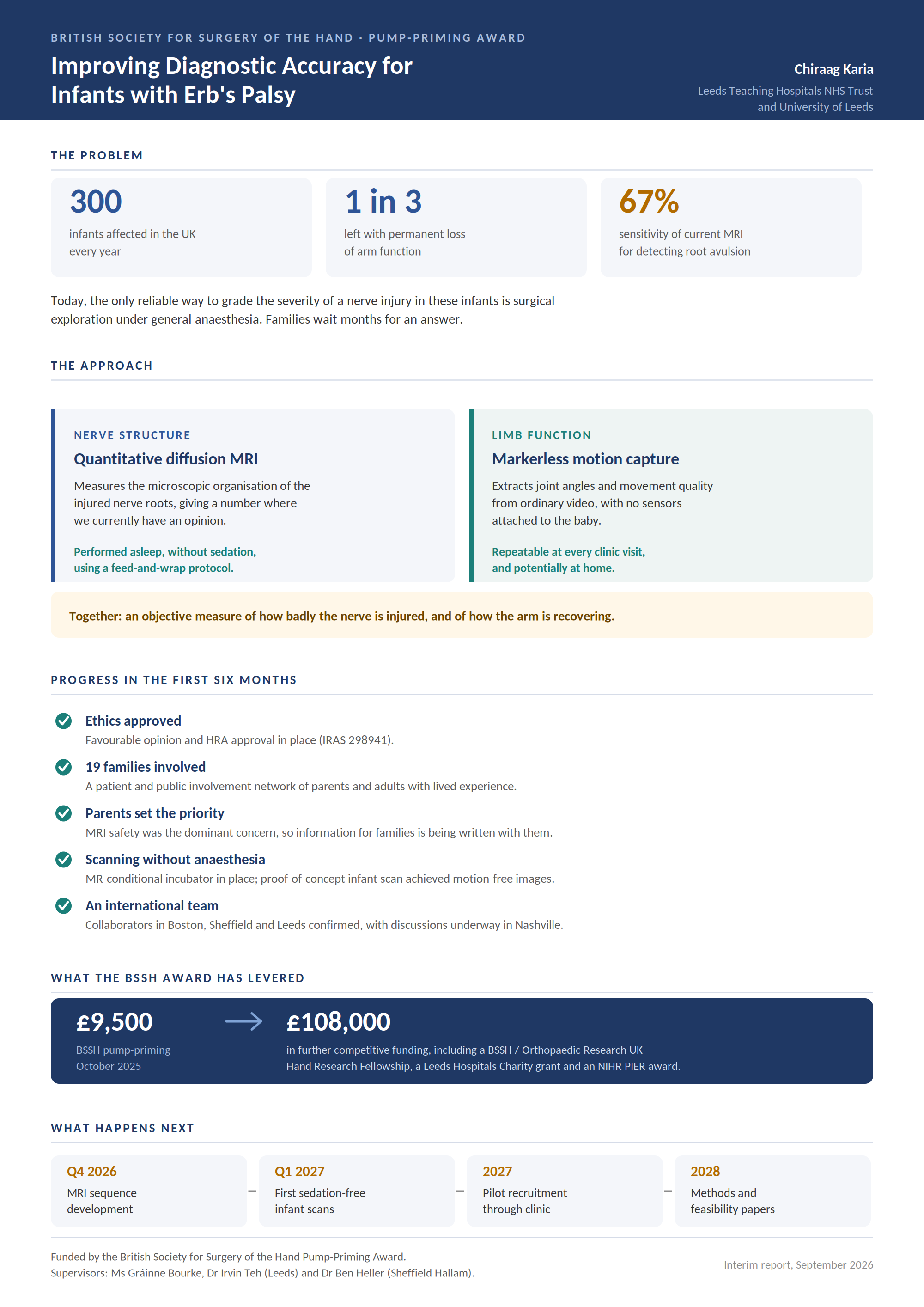
This report covers the first six months of the funded period, January to June 2026. The foundations of this project are considerably stronger than anticipated at the point of award, while the imaging workstream is behind schedule.
Amended ethical approval is in place, the sedation-free scanning infrastructure is established, an active patient and public involvement network of nineteen families has been built, and the international collaborations needed to deliver the work have been secured. The pump-priming funding has also levered three further competitive awards totalling approximately £108,000, including a BSSH/Orthopaedic Research UK Hand Research Fellowship, and has converted a ten-infant imaging pilot into a doctoral-scale research programme.
Sequence development and infant scanning have not yet begun. The reason is set out in section 5, together with the recovery plan and a request for an extension.
2. Progress against the funded objectives
|
Funded objective
|
Status
|
Commentary
|
|
Ethics and research governance approvals
|
Complete
|
Favourable opinion and HRA approval in place under IRAS 298941.
|
|
Establish patient and public involvement for the imaging pathway
|
Exceeded
|
Network grown to 19 families and adults with lived experience. Focus groups held; participant materials now being co-produced.
|
|
Secure sedation-free imaging infrastructure
|
Complete
|
MR-conditional incubator with high-density coils available at the Advanced Imaging Centre; feed-and-wrap pathway established with Dr Malenka Bissell.
|
|
Optimise a motion-tolerant DTI sequence for the infant plexus
|
Behind schedule
|
Physics work programmed to begin in Q4 2026 on return to Leeds. See section 5.
|
|
Pilot infant DTI scanning
|
Not yet started
|
First development scans planned for Q4 2026, with pilot recruitment through 2027.
|
|
Follow-on funding and dissemination
|
Exceeded
|
Three further competitive awards secured. See section 4.
|
3. Work delivered in the reporting period
Research governance. Ethical and HRA approvals are in place under IRAS 298941, covering diffusion imaging of the infant brachial plexus at Leeds Teaching Hospitals NHS Trust. Data management and information governance arrangements have been agreed with the Trust and the University.
Patient and public involvement. This has been the principal area of delivery. The network now comprises nineteen families and adults with lived experience, recruited through the specialist clinic, the Erb's Palsy Group and digital outreach, and co-led with Joanna Burdon, senior occupational therapist. Focus groups identified the safety of MRI in infants as the dominant parental concern, well ahead of scan duration or travel burden. Participant information sheets are consequently being co-produced with families to address this directly, using the Public Involvement Impact Assessment Framework to evaluate the influence of their input. This finding has already changed the design of the recruitment pathway and is, in itself, a useful output for the wider field.
Imaging infrastructure. Access to an MR-conditional incubator with high-density neonatal coils is confirmed at the Leeds Advanced Imaging Centre, and a feed-and-wrap pathway has been established with Dr Malenka Bissell. A proof-of-concept sedation-free infant scan has been performed, demonstrating that motion-free images can be obtained without general anaesthesia.
Collaborations. Professor Andrea Bauer (Boston Children's Hospital), who led the NAPTIME study, is confirmed as an international collaborator on the imaging protocol. Discussions are underway with Dr Kurt Schilling (Vanderbilt) on spinal cord and brain imaging as a methodological contingency. Dr Ben Heller (Sheffield Hallam) leads the movement science component, and Dr Irvin Teh provides MRI physics supervision in Leeds.
Scope. Following the involvement work and discussion with the supervisory team, a markerless computer vision workstream has been added, allowing limb function to be quantified serially from standard video without sensors. Nerve structure and limb function can therefore be measured together, which is the basis on which the follow-on funding has been awarded.
4. Funding levered by the BSSH award
The BSSH pump-priming grant was the first peer-reviewed award made to this project by a surgical specialty body, and was cited in each subsequent application. Together with HealthTech Leeds seed funding of £8,000 secured shortly beforehand, it has since levered:
- £9,224, Leeds Hospitals Charity Innovation Grant, supporting the motion capture workstream
- £710, NIHR Research Support Service PIER award, supporting public involvement
- £98,446, BSSH/Orthopaedic Research UK Hand Research Fellowship over 24 months, awarded September 2026 following a unanimous panel recommendation
Applications for doctoral funding to the NIHR, Action Medical Research and Forward North are in preparation or under review, with decisions expected during 2027.
5. Delay, explanation and recovery plan
Development of the motion-tolerant DTI sequence and the first infant scans have not progressed as planned. I was rotated to a clinical placement outside of Leeds for the majority of the funded period, which removed day-to-day access to the Advanced Imaging Centre and the MRI physics team on whom the sequence work depends. Rather than attempt the physics work remotely and risk a poor-quality protocol, the decision was taken to complete the governance, involvement and collaborative groundwork first.
I return to Leeds in October 2026 in a post with protected academic time, and the BSSH/ORUK fellowship provides 0.5 FTE from the start of that award. The recovery plan is:
- Sequence development sessions with the MRI physics team, beginning Q4 2026
- First sedation-free development scans on the optimised protocol, Q4 2026 to Q1 2027
- Pilot infant recruitment through the specialist clinic from Q1 2027
6. Outputs and dissemination
Work from the group underpinning this award and published during the period:
- Hardie CM, Karia CT, Wade RG, Kraft JK, Bains R, Bourke G. Magnetic resonance imaging for detecting root avulsions in brachial plexus birth injuries. J Hand Surg Eur Vol. 2026 May;51(5):589-596.
- Karia CT, Koroma P, Nelson-Ashley S, Olszewska P, Bourke G. The Utility of Magnetic Resonance Imaging of the Spinal Cord in Paediatric Transverse Myelitis: A Systematic Review. Multiple Sclerosis and Related Disorders. To Be Confirmed
The first of these established that conventional 3T MRI has a sensitivity of only 67% for root avulsion against surgical findings in our unit, which is the diagnostic gap this award was made to address.
Planned outputs from the funded work are a methods paper on sedation-free diffusion imaging of the infant plexus, a report of the involvement findings on parental acceptability of infant MRI, and presentation at the BSSH Scientific Meeting. The Journal of Hand Surgery (European Volume) is the intended primary outlet.
Chiraag Karia
NIHR Academic Clinical Fellow and ST5, Plastic and Reconstructive Surgery
Leeds Teaching Hospitals NHS Trust and University of Leeds